Let’s get transparent with x-rays.

Dr. Brady wants to provide the best possible dental care to our patients, but a visual examination doesn’t tell him everything he needs to know. Thanks to dental X-rays, Dr. Brady can accurately diagnose and treat dental problems early before they become more serious. And if after examining your mouth and reviewing these images, Dr. Brady finds no cavities or growth issues, you can rest assured he has seen the whole picture.

Dental X-rays are a useful diagnostic tool when helping your dentist detect damage and disease not visible during a regular dental exam. How often X-rays should be taken depends on your present oral health, your age, your risk for disease, and any signs and symptoms of oral disease. For example, children may require X-rays more often than adults because their teeth and jaws are still developing and their teeth are more likely to be affected by tooth decay than those of adults. Your dentist will review your history, examine your mouth and then decide whether or not you need X-rays.

If you are a new patient, Dr. Brady will recommend X-rays to determine the present status of your oral health and have a baseline to help identify changes that may occur later. A new set of X-rays may be needed to help Dr. Brady detect any new cavities, determine the status of your gum health or evaluate the growth and development of your teeth.

Dental X-ray exams are safe. They require very low levels of radiation exposure, which makes the risk of potentially harmful effects very small. Dental X-ray tools and techniques are designed to limit the body’s exposure to radiation and every precaution is taken to ensure that radiation exposure is As Low As Reasonable Achievable (the ALARA principle). A leaded apron minimizes exposure to the abdomen and may be used when it will not interfere with acquisition of the dental radiograph. Also, a leaded thyroid collar can protect the thyroid from radiation, and should also be used whenever possible. The use of a leaded thyroid collar is recommended for women of childbearing age, pregnant women and children.

X-rays are divided into two main categories, intraoral and extraoral. With intraoral X-rays, the X-ray film is inside the mouth. With extraoral X-rays, the film is outside the mouth.


Intraoral Radiographs

Intraoral X-rays are the most common type. They give a high level of detail. These X-rays allow Dr. Brady to: Find cavities Look at the tooth roots Check the health of the bony area around the tooth See the status of developing teeth Otherwise monitor good tooth health Otherwise monitor good tooth health

Bite-wing X-rays

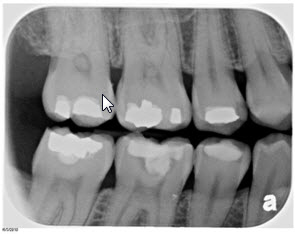
Highlight the crowns of the back teeth. Dentists take one or two bite-wing X-rays on each side of the mouth. Each X-ray shows the upper and lower molars (back teeth) and bicuspids (teeth in front of the molars). These X-rays are called "bite-wings" because you bite down on a wing-shaped device that holds the film in place while the X-ray is taken. These X-rays help dentists find decay between back teeth.

Periapical X-rays

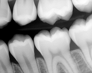
Highlight only one or two teeth at a time. A periapical X-ray looks similar to a bite-wing X-ray. However, it shows the entire length of each tooth, from crown to root. Depending on your oral health and dental history your dentist may recommend an x-ray.

Full mouth X-rays, or FMX

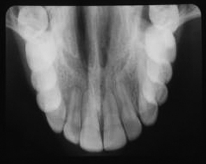
This includes every tooth, from crown to root to supporting structures. They are X-rayed using both bitewing and periapical radiographs.

Occlusal X-rays

are larger than most X-rays. They highlight tooth development and placement in children. Each X-ray shows nearly the full arch of teeth in either the upper or lower jaw.

Extraoral Radiographs

Extraoral X-rays are made with the film outside the mouth. These can be considered the “big picture” X-rays. They show teeth, but they also provide information on the jaw and skull. Extraoral radiographs are used to:

  • Keep track of growth and development
  • Look at the status of impacted teeth
  • Examine the relationships between teeth and jaws
  • Examine the bones of the face
Extraoral X-rays are less detailed than intraoral X-rays. For this reason, they are usually not used for detecting cavities or flaws in individual teeth


Panoramic X-rays

show the entire mouth on a single X-ray. They include all teeth on both upper and lower jaws. This type of X-ray requires a special machine. The tube head that emits the X-rays circles behind your head while the film circles across the front. That way, the full, broad view of the jaws is captured on one film. Because the machine moves in a set path, you have to be positioned carefully. Devices attached to the X-ray machine hold your head and jaw in place. All this may look and feel intimidating, but the process is very safe. It often uses less radiation than intraoral X-rays.

Digital radiographs

are one of the newest X-ray techniques. Standard X-ray film is replaced with a flat electronic pad or sensor. The image goes into a computer, where it can be viewed on a screen, stored or printed out. Digital X-rays taken at different times can be compared using a process that highlights differences between the images. Tiny changes therefore can be caught earlier. Used properly, digital X-rays use about half the radiation of conventional film.